Return to flip book view
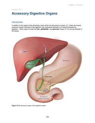
Chapter 15: Digestion 524 Lesson 15.2 Accessory Digestive Organs Introduction In addition to the organs of the alimentary canal, which are discussed in Lesson 15.1, there are several accessory organs important to the digestion and subsequent absorption of nutrients liberated by digestion. These organs include the liver, gallbladder, and pancreas (Figure 15.10) and are detailed in this lesson. Figure 15.10 Accessory organs to the digestive system.
Chapter 15: Digestion 525 15.2.01 Liver The liver is located in the upper right quadrant of the abdominal cavity, just below the diaphragm, and performs various functions (Figure 15.11). Figure 15.11 The liver is an accessory digestive organ. Prior to food consumption and the initiation of digestion, the liver plays an essential role in whole-body glucose homeostasis, supplying glucose to the blood for uptake by the tissues of the body. Upon food ingestion, concentrations of small molecules derived from carbohydrate, protein, and/or fat digestion increase in the blood. These molecules are delivered to the liver for processing. In addition, insulin is released by the pancreas (discussed in more detail in Concept 15.2.03), which signals the liver to take up glucose and reduce the production of glucose from gluconeogenesis and glycogenolysis. As the site of bile production, the liver is crucial for fat digestion (Figure 15.12). Bile (also discussed in Concept 15.2.02) is a solution released into the duodenum of the small intestine to help mechanically (ie, by physical, nonenzymatic means) digest lipids by breaking down large lipid globules into micelles (smaller droplets) during emulsification.
Chapter 15: Digestion 526 Figure 15.12 The liver is involved in bile production and release. The liver is also an important organ in the breakdown and detoxification of many drugs and waste products. Drugs are exogenous compounds that may have toxic effects when present in the body at elevated concentrations. The body produces endogenous waste products (eg, bilirubin, ammonia) that must be modified in the liver to avoid adverse effects. Macromolecules such as plasma proteins (eg, clotting factors and albumin), fats, ketone bodies, and cholesterol are produced by liver cells. Additionally, the liver stores several molecules (eg, glycogen), minerals (eg, iron), and vitamins. 15.2.02 Gallbladder The gallbladder is an accessory digestive organ. It does not synthesize molecules (eg, digestive compounds, enzymes); rather, the gallbladder is the storage reservoir for bile produced by the liver.
Chapter 15: Digestion 527 Bile is an alkaline fluid that facilitates fat digestion. Cholesterol, bile acids, and bile pigments (eg, bilirubin) are contained within bile. Before bile acids are released from the liver, they are conjugated with additional compounds to form bile salts, which are amphipathic (ie, containing hydrophobic and hydrophilic regions), a property essential for fat digestion. On a molecular level, hydrophobic regions of bile salts associate with fat globules, whereas hydrophilic regions associate with the aqueous environment. This allows bile salts to act as detergents and break down large lipid globules into smaller spherical structures called micelles (Figure 15.13). The formation of small micelles from larger lipid globules serves to increase lipid surface area for hydrolysis by lipases. Figure 15.13 Emulsification of lipids by bile salts. In the duodenum of the small intestine, the presence of meal-derived fats within chyme and the acidity of chyme itself stimulate bile release from both the liver and the gallbladder. Bile secretion into the duodenum promotes the neutralization of chyme and the physical digestion of fats (ie, emulsification). Emulsification is an example of mechanical digestion, a nonenzymatic process that physically breaks down food particles into smaller pieces. 15.2.03 Pancreas The pancreas, composed of various cell types (eg, alpha cells, beta cells, delta cells), has paracrine, exocrine, and endocrine functions. Cells with paracrine function secrete substances that exert effects on neighboring cells, and cells with exocrine function secrete substances (eg, saliva, sweat, enzymes) through a duct and onto an epithelial surface. In comparison, cells with endocrine function secrete hormones into the bloodstream to cause an effect in a different part of the body.
Chapter 15: Digestion 528 Endocrine cells of the pancreas secrete insulin and glucagon, which are hormones involved in the regulation of blood glucose, into the bloodstream (Figure 15.14). Exocrine cells of the pancreas secrete digestive enzymes and bicarbonate into the small intestine to assist in digestive processes and to neutralize the acidity of chyme. Figure 15.14 Involvement of the pancreas in the regulation of blood glucose.
Chapter 15: Digestion 529 The control of blood glucose is important in the maintenance of homeostasis. Food ingestion increases the blood glucose level and stimulates pancreatic beta cells to release insulin. Insulin is a glucose-regulating hormone that decreases the concentration of glucose in the blood. Insulin regulates glucose concentration in the blood by promoting glucose uptake by tissues (eg, adipose, muscle) and decreasing liver glucose production (ie, via gluconeogenesis, glycogenolysis), while at the same time promoting glucose storage in glycogen molecules (ie, glycogenesis). Insulin sensitivity refers to the biological response elicited by a fixed quantity of insulin. An organism is considered insulin-sensitive if only a minimal amount of insulin is needed to induce a reduction in blood glucose levels. In contrast, insulin-resistant organisms need substantially more insulin to stimulate cells to take up the same amount of glucose from the blood (Figure 15.15). Figure 15.15 Insulin resistance. Glucagon, a hormone released by alpha cells of the pancreas in response to low blood glucose levels, opposes the effects of insulin. Release of glucagon results in increased gluconeogenesis and glycogenolysis and decreased glycogenesis. Another hormone important to digestion is somatostatin, which is released by pancreatic delta cells. This hormone has a generalized inhibitory effect on digestive function and has been shown to suppress insulin and glucagon release. Figure 15.16 summarizes the cells of the pancreas and their products.
Chapter 15: Digestion 530 Figure 15.16 Cells of the pancreas and their products. The exocrine pancreas assists in chemical digestion, which is carried out by acids and enzymes and involves the cleavage of chemical bonds within macronutrients to form simpler compounds that can be absorbed. The formation of these compounds occurs through the secretion of enzymes into the pancreatic duct, which empties into the duodenum. These enzymes aid in the digestion of chyme in the lumen of the small intestine. Enzymes secreted by the pancreas include pancreatic lipase, which acts to chemically digest lipids, and pancreatic amylase, which facilitates polysaccharide hydrolysis to form disaccharides. The pancreas also secretes proteolytic digestive enzymes (eg, trypsinogen, chymotrypsinogen). A duodenal enzyme, enteropeptidase, converts the zymogen trypsinogen to its active form, trypsin. Trypsin activates other pancreatic zymogens (eg, converts chymotrypsinogen to chymotrypsin) and functions in continued peptide digestion.
Chapter 15: Digestion 531 Concept Check 15.2 Complete the table to match each accessory digestive organ (ie, “gallbladder,” “pancreas,” and “liver”) with its function. Each organ may be used more than once, and some functions may have more than one related organ. Accessory digestive organ(s) Function Lipid digestion Secretion of trypsinogen and chymotrypsinogen Production of bile Detoxification of drugs and waste products Secretion of somatostatin Glucose homeostasis Storage of bile Solution Note: The appendix contains the answer.